[vc_row][vc_column width=”1/3″][vc_column_text][mega_main_menu location=”mega_main_sidebar_menu”][/vc_column_text][/vc_column][vc_column width=”2/3″][et_parent tab_style=”linebox” tab_animation=”bounceInUp” color_tab_txt=”#064d70″ color_act_txt=”#dd3333″ color_act_bg=”#e0e0e0″ color_content_bg=”#ffffff”][et_single icon=”no-icon” tab_title=”
 It is essential to know how a "suspicious" nevus is evaluated: Nevus are roughly evaluated based on the ABCD algorithm, where A corresponds to asymmetry (asymmetry, one half is not similar to the other half) B corresponds to border (irregular, or vaguely outlined borders of the lesion) C corresponds to the color (variegated color that includes shades of brown, black, white, red or blue) D corresponds to the diameter (diameter greater than 6 millimeters or growing in size nevus). The patient's self-examination is considered very important. If a patient notices changes in a mole, they should see their Dermatologist for an examination. Noticeable changes in a nevus are considered: -Sudden bleeding (without the patient remembering any injury) -Change in the sensibility of the nevus (stinging, itching-itching) -Changes in color and mainly appearance of color variety in the same lesion. -Changes in shape, mainly the appearance of pseudopods (protrusions, small feet) in the periphery of the lesion -Changes in the nevus and/or in the surrounding area of the skin: erythema (redness), edema (swelling), ulcer (wound that does not close) Finally, people with burdened family history (melanoma) or numerous moles, or large familial (present from birth) moles should be preventively examined by a dermatologist at least once a year. In these patients, it makes sense to save the dermatoscopic images for comparison at the next check-up, a process called mapping.
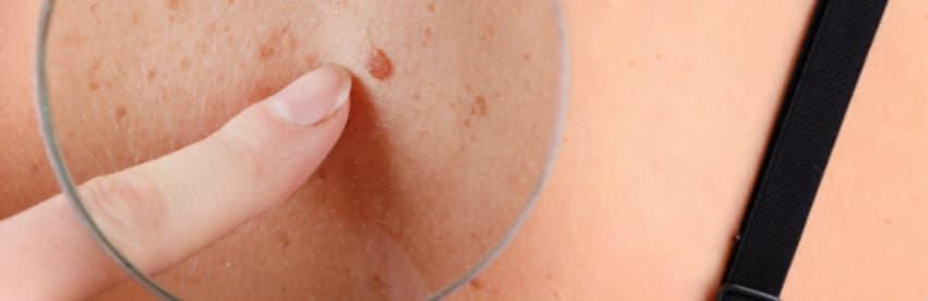 "Dermoscopy is a weapon that dermatologists have had for the past decade to very effectively examine the form and internal structure of a mole, a mole. With dermoscopy, we see the inside of the olive, we see its internal symmetry, we see its internal shapes, so if we see something worrying, we put the patient on a regular follow-up, in a mapping which depicts all his olives in the body, or we recommend surgical removal suspicious olive. This way we can treat a possibly suspicious lesion very effectively and avoid its progression, its transformation into a skin cancer which is something very serious. The procedure is easy, it is completely painless for the patient, it does not take long and it offers us very important security."[/et_single][et_single icon=”no-icon” tab_title=”
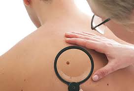
Dermatoscopy
Dermoscopy is the examination of the skin with the help of a dermoscope. It is a non-invasive, diagnostic technique that magnifies skin lesions to such an extent that the diversity in their color, internal architecture and morphological characteristics of the lesion becomes visible, elements that cannot be seen with the naked eye or with normal magnification of common magnifying lenses. With proper training and experience, dermoscopy improves the diagnosis of pigmented and non-pigmented, benign and malignant skin lesions, with the main benefit being its contribution to the early diagnosis of melanoma. In addition to the wide appeal of dermoscopy in the examination of moles (skin olives), its use also extends to other skin diseases such as hemangiomas, seborrheic and actinic hyperkeratoses, epitheliomas of the skin (basal cell, squamous cell).
It is essential to know how a "suspicious" nevus is evaluated: Nevus are roughly evaluated based on the ABCD algorithm, where A corresponds to asymmetry (asymmetry, one half is not similar to the other half) B corresponds to border (irregular, or vaguely outlined borders of the lesion) C corresponds to the color (variegated color that includes shades of brown, black, white, red or blue) D corresponds to the diameter (diameter greater than 6 millimeters or growing in size nevus). The patient's self-examination is considered very important. If a patient notices changes in a mole, they should see their Dermatologist for an examination. Noticeable changes in a nevus are considered: -Sudden bleeding (without the patient remembering any injury) -Change in the sensibility of the nevus (stinging, itching-itching) -Changes in color and mainly appearance of color variety in the same lesion. -Changes in shape, mainly the appearance of pseudopods (protrusions, small feet) in the periphery of the lesion -Changes in the nevus and/or in the surrounding area of the skin: erythema (redness), edema (swelling), ulcer (wound that does not close) Finally, people with burdened family history (melanoma) or numerous moles, or large familial (present from birth) moles should be preventively examined by a dermatologist at least once a year. In these patients, it makes sense to save the dermatoscopic images for comparison at the next check-up, a process called mapping.
"Dermoscopy is a weapon that dermatologists have had for the past decade to very effectively examine the form and internal structure of a mole, a mole. With dermoscopy, we see the inside of the olive, we see its internal symmetry, we see its internal shapes, so if we see something worrying, we put the patient on a regular follow-up, in a mapping which depicts all his olives in the body, or we recommend surgical removal suspicious olive. This way we can treat a possibly suspicious lesion very effectively and avoid its progression, its transformation into a skin cancer which is something very serious. The procedure is easy, it is completely painless for the patient, it does not take long and it offers us very important security."[/et_single][et_single icon=”no-icon” tab_title=”
Video
“] Edit this text with your custom content. [/et_single][et_single icon=”no-icon” tab_title=”Appointment
“][gravityform id=”3″ name=”Diagnosis appointment” title=”false” ajax=”false”][/et_single][/et_parent][/vc_column][/vc_row]